Reilly Carbon Monoxide Poisoning

Home » Veterinarians » Case Studies » Reilly

History

Reilly, a four-year-old miniature Goldendoodle, presented to Southeast Veterinary Neurology for acute neurologic signs, which began four days after being caught in a house fire. Prior to the house fire, she was a very healthy and happy dog.

Reilly Smiling
Happy Reilly

On the day of the fire, Reilly was admitted to an emergency animal hospital, where she was treated supportively for smoke inhalation and discharged a few days later. But the following day, she developed abnormal neurologic signs and returned to the hospital, where it was recommended she be taken to Southeast Veterinary Neurology of Jupiter for a consultation.

Examination

Upon examination, Reilly was alert, responsive, and fully ambulatory. She displayed no paresis (weakness) or ataxia (unsteady gait); only a constant, rhythmic spasm of her left thoracic limb (forelimb). She did stumble a couple of times while walking, and at one point, she lost her footing when she shook; her chin hitting the floor.

Although she was able to navigate around the room, cranial nerve assessment revealed an absent menace response in both eyes, an absent palpebral response from the outer corner of the eyes, and slightly reduced sensation on the inside of her nose. Additionally, proprioceptive (awareness of paw placement) deficits were noted on the left side of her body when she did not correct the left fore or hind paws if they were turned upside down. The remainder of the neurologic examination was unremarkable.

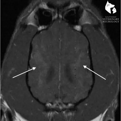
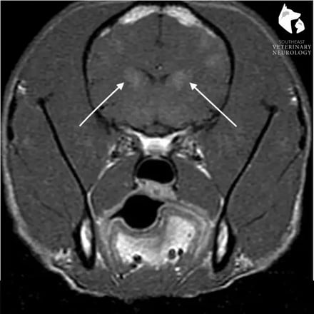
Based on the examination findings, Reilly’s neuroanatomic lesion localization was intracranial (in the brain), which raised concern for central nervous system inflammatory disease (meningoencephalitis); as it was also suspected that the abnormal movement of the left forelimb mentioned earlier was actually a manifestation of seizure activity.

Diagnosis

Chest radiographs and complete blood work were all normal, aside from elevated ALT (liver enzyme), which was most likely due to the smoke exposure. Finally, an MRI of Reilly’s brain revealed bilaterally symmetric contrast-enhancing lesions within the cerebral cortex and caudate nuclei, while cerebrospinal fluid analysis was normal. This was most consistent with a metabolic or toxic etiology. Therefore, the most likely cause was carbon monoxide toxicity to the brain from smoke inhalation.

Reilly MRI
MRI Of Reilly's Head

Treatment

Reilly was treated with both anti-seizure levetiracetam and a short course of anti-inflammatory prednisone. Her limb movements completely resolved, but she continued to have mild visual deficits in both eyes, and the owner reported that Reilly remained a bit “off” on the left forelimb. However, at her two-week recheck, she was much brighter, with a normal gait and steady footing.

Takeaway

Reilly’s case is a classic example of toxic insult to the brain. Although it has been reported in veterinary literature, toxic changes within the brain due to smoke inhalation are not commonly identified in veterinary patients because clinical signs are typically delayed from the time of incident. Reilly’s case teaches us the importance of keeping toxicity on the list of possible causes for neurologic signs. Fortunately, prognosis for recovery is good. For our sweet Reilly, only time will tell whether or not her deficits will fully resolve. What we can say is that her quality of life in the meantime is excellent, thanks to prompt treatment by the experts at Southeast Veterinary Neurology of Jupiter.