MRI for Pets
Home » Diagnostics and Treatment » MRI
Why does my pet need an MRI?
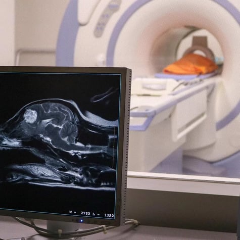
Magnetic Resonance Imaging (MRI) is a completely noninvasive, pain-free way of evaluating the internal structures of the body, and it is the advanced imaging modality of choice for examining soft tissues, including the brain and spinal cord.
One neurological symptom can have many different causes. Finding the exact cause such as tumors, strokes, meningitis, and intervertebral disc disease is important, so we can figure out the best treatment plan and gauge your pet's likelihood of recovery.
Most conditions affecting the brain or spinal cord are best tested for with an MRI. We may recommend an MRI of the head if a pet is showing symptoms of seizures, balance problems, or behavior changes. We may recommend an MRI of the neck or back if a pet is showing symptoms of weakness, pain, wobbly walking, or inability to walk.
We will only recommend an MRI if it is in your pet's best interest. Our vet neurologists carefully weigh the pros and cons of doing an MRI and discuss these with you in depth.
Why not just use X-rays (radiographs) or CT scans?

Radiographs and CT scans use X-rays to look at the internal structures of the body. X-rays are good for looking at the bones and lungs, because air is dark while bone and metal are white. However, they typically aren’t very effective for evaluating the soft tissues of the brain or spinal cord, because all tissues present as a similar shade of gray.
MRI, on the other hand can accentuate various types of tissues, providing much better tissue resolution. Therefore, MRIs can show many diseases that CT scans will miss.
Many neurological problems require MRI for a definitive diagnosis. By having an in-house CT scanner and the most powerful MRI for pets, we can save clients time and money by offering the most efficient and accurate testing possible.
How is an MRI performed?

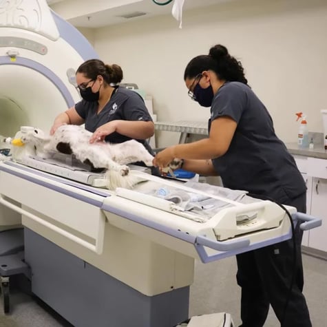
Using a strong magnet and specialized equipment, MRI devices are able to make a more comprehensive picture of the inside of a patient’s body.
Patients must remain completely still for the entire MRI, so general anesthesia is required. However, an advantage of choosing Southeast Veterinary Neurology (SEVN) is that our MRI works faster than less powerful MRIs, so pets do not need as much anesthesia. This makes the procedure much safer.
The patient will lie on a soft bed with blankets to stay warm during the scan. A veterinary nurse is always right by its side to ensure safety and monitor the patient closely during the procedure.
Why choose Southeast Veterinary Neurology for pet MRI?

We use a high-field 1.5 Tesla MRI, which is the best technology available in veterinary medicine. Compared to low-field MRI, our MRI provides images with more clarity in less time. This means a more accurate answer with less need for anesthesia. In fact, our vet neurologists routinely find abnormalities that other imaging services miss. Compare the MRIs below.
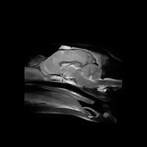
Low-field MRI of the brain performed at another facility.
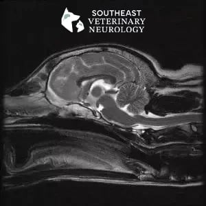
High-field MRI of the brain performed at Southeast Veterinary Neurology. Note the improved quality.
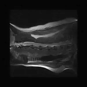
Low-field MRI of the neck performed at another facility.
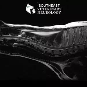
High-field MRI of the neck performed at Southeast Veterinary Neurology. Note the improved image quality.