First, is Lupa’s problem a neurological one? Absent postural reactions (“CP deficits”) are highly suggestive of a neurological problem.
Second, where is Lupa’s neurological problem? From a simplistic view, the facts that she is alert and that her cranial nerves are normal suggest that the problem is not the brain. Her spinal reflexes are normal, as evidenced by her ability to withdraw her legs when gently pinched. She is able to move her legs, but not strong enough to walk in all four legs. This is termed non-ambulatory tetraparesis. Since she is alert, with normal cranial nerves, normal reflexes, but all four legs are affected, her problem is in the cervical spinal cord.
Third, her problem is severe in that she is unable to walk, but is able to feel her legs.
Differential diagnoses include a slipped disc, a spinal tumor, a malformation (cyst, syringomyelia, atlantoaxial instability), meningitis/myelitis, infection, or other less likely cause.
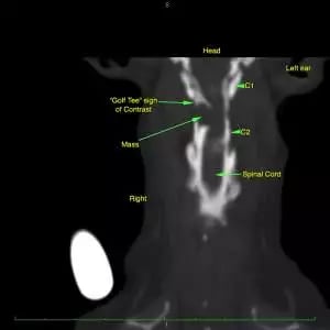
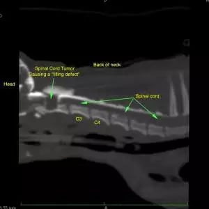
A complete blood count (CBC), chemistry panel, and thoracic radiographs were performed. A CT/myelogram was performed at another hospital and showed an abnormality at C1-C2. Remember, there are three main myelographic patterns:
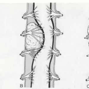
- Intramedullary: Inside the parenchyma of the spinal cord. The spinal cord may appear ‘swollen’ and the contrast columns deviate from each other.
- Intradural-extramedullary: Inside of the dura, but not inside the spinal cord itself. Contrast in the subarachnoid space flows up to the abnormality, and there may be a ‘filling defect’ within the subarachnoid space. A ‘golf-tee’ sign may be seen.
- Extradural: Outside of dura – the outermost layer of the meninges. The contrast column will typically ‘indent’ in towards the spinal cord.
Note the ‘golf-tee’ sign, which is classic for an intradural-extramedullary mass. Given the severity of her signs, the location of the presumed tumor and her small size, a poor prognosis was given at the first hospital and euthanasia was discussed.
Dr. Wong was consulted as a second opinion. A tumor was considered most likely. Common tumors in the intradural-extramedullary space include meningioma, peripheral nerve sheath tumor, or metastatic tumor such as hemangiosarcoma. The best scenario would be a meningioma. After discussing the pros and cons of surgery, the owners elected surgery. A laminectomy was performed at the caudal aspect of C1 and the cranial aspect of C2. A durotomy was performed and a large, grey-to-tan, firm-to-friable mass was present in the subarachnoid space. The mass was removed. She was treated with pain medications and the prednisone was tapered over the next two weeks. The mass was submitted for histopathological analysis and was a meningioma.