Luna Bell Syringomyelia

Home » Veterinarians » Case Studies » Luna Bell

Luna Bell, a 10 month-old female intact Chihuahua presented for evaluation of episodes of crying. She was adopted from Puerto Rico and has been overall healthy. She received her vaccinations in a timely fashion and has recently started heartworm preventative. She is fed a commercial diet, has a good appetite, and urinates and defecates normally. She does not have any coughing, sneezing, vomiting or diarrhea.

The owner reports that Luna Bell cries spontaneously. She is playful and wants to interact. There does not seem to be a rhyme or reason to when she cries. Sometimes she can be lying down and other times she is being active.

The neurological examination consists of evaluation of mentation, gait, postural reactions, spinal reflexes, cranial nerves, and palpation. It aims to determine IF the problem is neurological in origin, WHERE in the nervous system the problem is (e.g. the neuroanatomical localization) and HOW BAD the problem is. Using this information, the clinician can formulate a reasonable list of differential diagnoses and a diagnostic plan.

Mentation: Luna Bell is alert and responsive.

Gait and Posture: She is able to stand and walk, but is slightly ataxic in the thoracic limbs. She knuckles over in the thoracic limbs and sometimes seems to “lead" with her rear end. She carries her head somewhat low at times, but will shake her head like a normal dog.

Postural Reactions: Her postural reactions are delayed in all four limbs.

Spinal Reflexes: These are local reflex arcs. At Southeast Veterinary Neurology, we primarily test the patellar reflex, flexor withdrawal reflexes in all four limbs, cutaneous trunci reflex, and perineal reflex. These were all normal.

Is this patient’s problem neurological in origin?

Where is the problem?

What are your differential diagnoses?

Radiograph Pre Op

What do you see?

Luna Berger Mri Pre Op

MRI of Luna Bell’s head and neck

Luna Bell CT

Luna CT

Luna Bell’s examination indicates a neurological problem affecting the C1-C5 spinal cord. She is alert and responsive, has normal cranial nerves, has postural reaction deficits in all four limbs, but has normal spinal reflexes in all four limbs.

Possible causes include a malformation such as atlantoaxial instability, Chiari-like malformation and syringomyelia, meningitis/myelitis, infection, slipped disc (unlikely based on age), tumor (unlikely based on age) or other less likely cause.

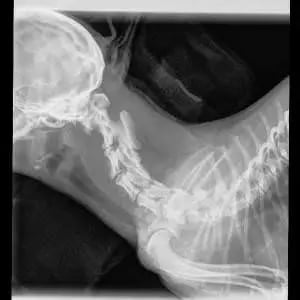
Radiographs of the cervical vertebral column were performed.

There is increased space between the dorsal aspect of C1 and the cranial/dorsal aspect of C2. There is subluxation at C2 relative to C1, with dorsal angulation of the caudal portion. Note that one cannot visualize the spinal cord nor the intervertebral discs. Additionally, there is an overlap of structures within the cervical region. Atlantoaxial instability and subluxation was diagnosed through the radiographs.

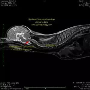
An MRI was performed to further define the abnormalities. Multiple congenital abnormalities can occur concurrently (e.g. synovial cyst associated with AA instability, etc). MRI shows these soft tissue abnormalities best and is the only way to diagnose Chiari-like malformation and syringomyelia.

MRI is superior for evaluating the soft tissues of the brain, spinal cord, meninges, and discs. Note the ‘kinking’ of the cranial cervical spinal cord where it is being compressed by the dorsal subluxation of C2 relative to C1.

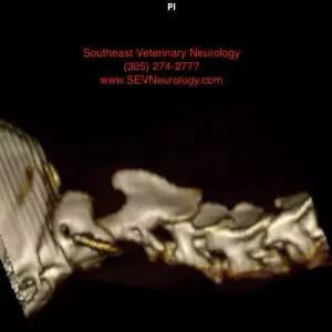
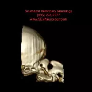
At Southeast Veterinary Neurology, we typically perform a CT scan prior to surgery. While MRI is the ideal way of imaging the spinal cord and brain, CT is excellent for evaluating the bone. The treatment of choice for atlantoaxial instability is surgical stabilization with screws and bone cement. Since AA instability is typically diagnosed in small dogs (sometimes less than 1kg), pre-operative planning for size and angle of the implants is indicated. Below is a 3-dimension reconstruction of Luna Bell’s cervical vertebral column and caudal skull. CT and MRI are able to be performed under the same anesthetic period at Southeast Veterinary Neurology.

At Southeast Veterinary Neurology, we have the ability to print 3D images of our CTs. Below is a 3D print of Luna Bell’s vertebral column. This can be used to help plan the surgery.

An atlantoaxial stabilization procedure was scheduled. Stabilization is provided by forming an arthrodesis at C1-2. There are several reported ways of providing stabilization. The preferred method at Southeast Veterinary Neurology is with multiple screws placed into C1 and C2 (+/- C3) fused with polymethylmethacrylate. The size and number of implants depend on the size of the patient. Below is a photograph from Luna Bell’s surgery. In the upper left, there is a video screen that shows a low-resolution version of what Dr. Wong is seeing through the operating microscope. On the screen, we are viewing the ventral aspect of the vertebral column. Cranial is toward the left of the screen and the left side of the dog is toward the top of the screen. Three 1.5mm titanium self-drilling screws have been placed into C1. Three screws have also been placed into C2. There are two in the cranial aspect of C2 and one on midline in the caudal aspect of C2. The articular cartilage at C1-2 is debrided with a #11 scalpel blade and house curette. In larger dogs, a small burr can be used to remove the articular cartilage. The joint is packed with bone graft. Polymethylmethacrylate is then placed over the screws to stabilize the construct.

Post-operatively, another CT scan was performed to ensure proper placement of the screws, excellent coverage of the screws by the bone cement and reduction of the subluxation. Here is a 3D reconstruct of the post-op CT. Notice that the subluxation is reduced and that there is excellent alignment. The screw placement was perfect with excellent bone purchase and angulation.

Post-operatively, Luna Bell was placed into a neck brace to limit her activity and stress on the arthrodesis. She is doing well and is recovering. She is walking well and has not had any evidence of pain.

Aa Sx Screw Placement

Luna's Screw Placement

Luna CT Post Treatment

Luna's CT Post Treatment

Luna Post Treatment

Luna Post Treatment