History
Bear, a 7-year-old neutered male Wheaten Terrier, presented to Southeast Veterinary Neurology (SEVN) in August of 2020 laterally recumbent and minimally responsive. Prior to presentation, Bear was hospitalized at a pet emergency center with dull mentation, nystagmus, and fever. Despite supportive care, he continued to decline and was referred for neurologic evaluation.
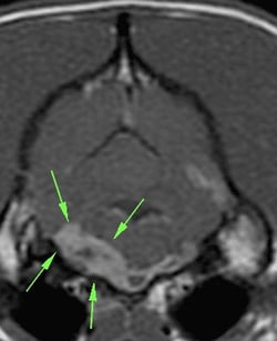
Transverse T1 postcontrast image at the level of the brainstem. The arrows are outlining the bulk of the mass along the left ventral aspect of the brainstem, but it extends to the right side.
Diagnosis
Transverse T1 postcontrast image at the level of the brainstem. The arrows are outlining the bulk of the mass along the left ventral aspect of the brainstem, but it extends to the right side.
Bear’s X-rays indicated bilateral cranioventral alveolar infiltrate (fluid in the lungs), worse on the left side than the right, consistent with bronchopneumonia.
In order to evaluate his brain, MRI was performed by SEVN. The MRI showed a mass along the bottom of his brainstem, which was causing significant swelling within the brainstem. The swelling was causing an obstruction of the flow of cerebrospinal fluid (CSF), called obstructive hydrocephalus. Hydrocephalus is a buildup of CSF in the brain. In Bear’s case, this was causing an elevation in intracranial pressure (pressure in the skull), which then affects perfusion of the brain (blood flow to the brain).
His CSF analysis showed a marked neutrophilic pleocytosis (increase in white blood cells, specifically neutrophils). This can be seen with tumors called meningiomas, though can also be seen with infectious diseases.
Treatment
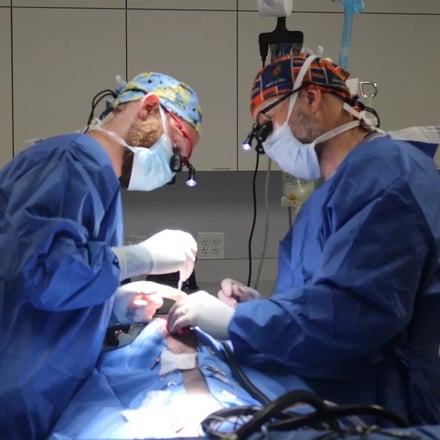
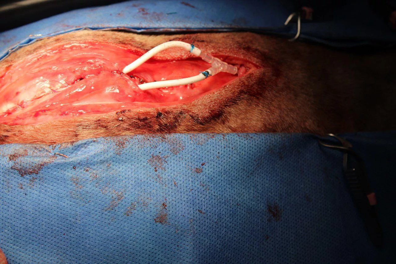
SEVN surgically placed ventriculosubcutaneous shunts (tubes that redirect CSF to another part of the body for reabsorption) into Bear’s lateral ventricles (chambers in the brain that contain CSF) to allow CSF to drain into the space under the skin at the top of his neck. This was a temporary solution to immediately alleviate intracranial pressure in order to stabilize him and formulate a treatment plan for the mass. He also prescribed steroids to reduce the inflammation within the brainstem caused by the mass.
Bear recovered well after surgery, and was able to walk out the door at the time of discharge, three days later. After discharge, infectious disease testing of his CSF came back negative for all diseases tested, which suggested the mass in his brainstem was cancerous. Bear began radiation therapy at a cancer center. His condition dramatically improved following treatment, and he continued to do great over the next few months.

Transverse T1 postcontrast image at the level of the brainstem, 17 months later. The mass is much reduced in size. The fourth ventricle is dilated, and the ventricular catheters are visible within the soft tissues dorsal to the skull.
Transverse T1 postcontrast image at the level of the brainstem, 17 months later. The mass is much reduced in size. The fourth ventricle is dilated, and the ventricular catheters are visible within the soft tissues dorsal to the skull.
Unfortunately, by January, he was presenting to SEVN again for disorientation, incoordination, and collapse. A repeat MRI confirmed that his brainstem mass was, in fact, shrinking. However, the MRI also identified enlargement of the entire ventricular system, meaning CSF was building up in the brain again. Since no obvious obstruction was observed, the cause of hydrocephalus was unclear. This implied that the temporary shunts had failed, but that shunts were still necessary.
So this time, Bear was treated with a more permanent solution. In a second surgery, SEVN placed ventriculoperitoneal shunts to divert excess CSF from his brain into his abdomen. Additionally, a suboccipital decompression (a procedure removing part of the back of the skull) was performed to remove any obstruction that may have been restricting the flow of CSF.
During surgery, very little spinal fluid was produced upon initially opening the meninges (membranes that cover and protect the brain and spinal cord). A thin layer of abnormal tissue was discovered at the base of the cerebellum (back part of brain) at the cerebellomedullary cistern. This may have been scar tissue from the mass or radiation, and upon its removal, the spinal fluid was readily flowing through the area.
This did the trick, and Bear has been doing fantastic ever since. At all of his subsequent rechecks, he was bright and alert with normal mentation, ambulatory with no signs of weakness, and not in any pain. His only remaining clinical sign was a subtle menace response deficit in his right eye. Other than that, he has been neurologically normal and back to his normal happy self at home.