Baby Brain Tumor, Meningioma

Baby, a 16-year-old castrated male Domestic Shorthair presented to Southeast Veterinary Neurology (SEVN) with a one-year history of ear infections. He had been treated with prednisone and ear medications and seemed to improve; however, signs would recur. He was never off balance, but just seemed dull and reluctant to jump onto the furniture and countertops. The night prior to presentation to SEVN, he became very lethargic and was unresponsive for about eight hours.

On examination, Baby was dull and distant. He is unable to stand and walk on his own. When supported to stand, he crouches low, takes a couple of steps and lays down or falls. He seems to scuff the thoracic limbs when trying to walk. He has a tendency to fall to the left, but there is no evidence of head tilt or abnormal eye movements. When ‘hopped’ on one leg, he does not accurately hop on any of the limbs, however, the left thoracic limb is the worst. He does not respond appropriately when a hand is brought toward his eye.

Baby's Examination

His examination suggests a problem affecting the cerebral cortex, slightly more towards the right side. The possible causes included a degenerative brain condition, low blood sugar, brain tumor, nutritional deficiency, brain infection or other less likely cause.

Blood tests including a CBC, chemistry panel, T4, and urinalysis were unremarkable. Thoracic and abdominal radiographs were unremarkable. Magnetic resonance imaging (MRI) was performed.

Baby MRI

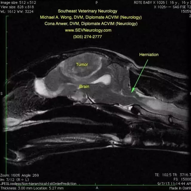
This first image is a sagittal, T2-weighted MRI of Baby’s head. The nose is toward the left and the top of the head is toward the top of the image. Note the evidence of mass effect--the brain tissue is being pushed down by the mass. The cerebellum is normally round, but you can see how it is being squashed toward the right and that there is herniation (arrow) of the cerebellum. A computed tomography (CT) scan would not show these changes.

Baby Mri

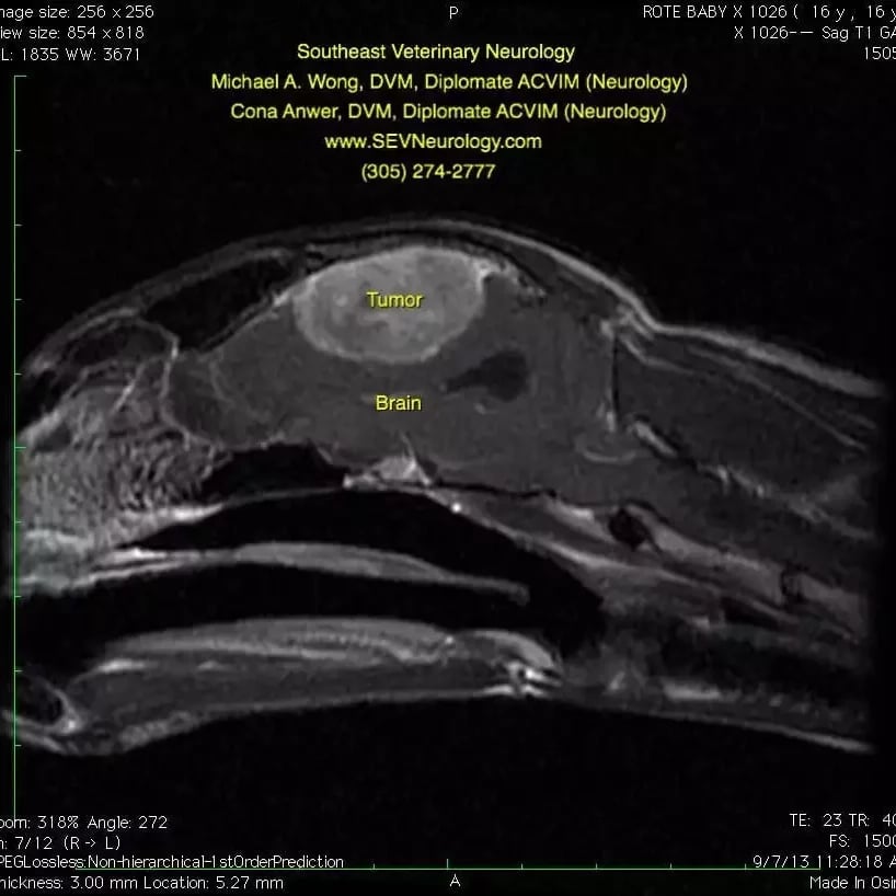
This second image is a T1 weighted sagittal image of Baby’s head. Contrast agent has been administered. The tumor enhances with the contrast agent. Note that the tumor appears to grow from the outside and push inward on the brain. Furthermore, there is a ‘tail’ at the cranial/dorsal and caudal/dorsal aspects of the mass. This is called a ‘dural tail’ sign. While a definitive diagnosis cannot be obtained without a biopsy, the most common cause of a large, contrast-enhancing mass with broad-based dural attachment in a 16-year-old cat is a meningioma.
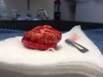
Baby Maningioma

Surgery

After discussing the potential risks and benefits of surgery, the owner elected to have the brain tumor removed. Dr. Wong removed the tumor with surgery. The mass was firm and adhered to the bones of the head. A photo of the tumor after surgery is shown here.

Post Surgery

Baby was hospitalized for three days after surgery for pain control, nutritional support, and nursing care. The biopsy showed that the mass was a meningioma. Meningiomas tend to be very slow-growing brain tumors that are often cured by surgery. Here is a video of Baby at his two-week follow-up. You can see that he is much more alert and happy. He lived to be almost 20.