Max
Brain Tumor
Home » Case Studies » Max
Max, a 10-year-old intact male Boxer, presented to Southeast Veterinary Neurology for being off balance. Max belongs to a local veterinarian who noticed that at times Max was uncoordinated. Sometimes Max would be completely normal and, at other times, he would stop suddenly and not move. He had a subtle head tilt toward the left.
Max was otherwise healthy, with a good appetite. His physical examination was normal, with no heart murmurs and minimal waxy debris in the right ear canal. Max’s owner, a veterinarian, performed chest X-rays, blood work including a CBC, chemistry panel, and thyroid profile. The thyroid profile showed a mildly low T4 with a normal free T4. He was otherwise not clinically hypothyroid. Chest X-rays were normal.
Max was already on antibiotics for a recently-diagnosed urinary tract infection.
On examination at Southeast Veterinary Neurology, Max was alert, with a subtle head tilt toward the left. Postural reactions (knowledge of where his feet are) were normal. He was able to walk without any evidence of weakness (paresis) or incoordination (ataxia). The rest of his cranial nerves were normal.
Initially, an MRI was not performed with the hope that the antibiotics he was on would treat a possible ear infection. Max was treated for another week or two but began having more frequent episodes of being off balance. An MRI of Max’s head was performed to find the cause of his balance problems.

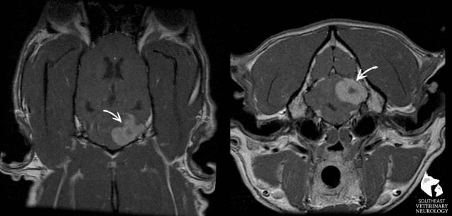
This is Max’s MRI. On the left is a coronal view with Max’s nose toward the top of the picture. The picture on the right is a transverse image of his head at the level of the ear canals. Note the large, round white mass on the right side of his cerebellum. This mass was suspected to be a meningioma. Meningiomas tend to be benign, slower-growing tumors of the coverings of the brain. They are typically diagnosed in older, long-nosed breeds.
Several treatment options were discussed, including surgery to remove the tumor and confirm what type of tumor it was, radiation to try to shrink the tumor or medications such as prednisone. The owners elected surgery, which was planned for the following week.
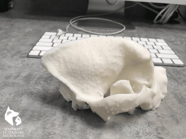
A 3-dimensional model was made of Max’s skull to plan for surgery.


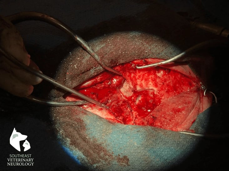
The following are several images from Max's brain surgery.
The first image is a photo of the craniectomy. A high-speed burr was used to make a window in the skull. The main concern with Max’s surgery was that the tumor was behind the transverse sinus, which is one of the main ways that blood is drained from the brain. The transverse sinus was identified and occluded to prevent bleeding.
The following is a video of the tumor removal. The tumor is removed gently while minimizing handling of the normal brain tissue. Most of the tumor was removed in this manner. A specialized instrument, called an ultrasonic aspirator, was then used to remove the residual tumor.
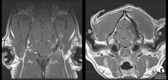
An MRI was performed immediately after surgery to assess for any complications such as bleeding or swelling of the brain. Additionally, the MRI is used to determine if any tumor was left behind. Typically, some tumor is left behind since “margins" of normal tissue (meaning normal brain) cannot be removed.
Note that the vast majority of the tumor is removed when compared to the pre-surgery MRI.

Max recovered well from anesthesia and was hospitalized for 3-4 days after surgery. Here is a video of him 2-3 days after surgery catching treats. As you can see, he is very comfortable and is very coordinated.
Max’s tumor was sent to the lab to determine the type of cancer and whether it was aggressive or benign. Max’s tumor was indeed a meningioma. The mass was also sent for production of an anti-tumor vaccine that is aimed to kill remaining tumor cells and improve his quality and quantity of life.
Max is currently doing very well and is back to normal at home.
To learn more about brain tumors in dogs and cats visit this link or call an expert in neurology at Southeast Veterinary Neurology. We have locations in Miami, Jupiter, Boynton Beach and Virginia Beach.