Skip to content

Koda
Bacterial Meningitis

Home » Case Studies » Koda

History

Koda, a 4-week-old intact female Goldendoodle, presented to Southeast Veterinary Neurology (SEVN) in March of 2021 for inability to walk.

Koda was purchased from a breeder the previous day. She suddenly developed a head tilt to the left and was taken to an emergency facility where she was hospitalized.

However, she worsened overnight and became unable to walk. Koda was referred to SEVN the next morning.

koda-exam

Examination

koda-exam

Upon examination, Koda was quiet, but responsive. She was non-ambulatory (unable to walk), but if supported, she displayed a vestibular-quality ataxia (incoordination), falling toward the left.

Cranial nerve assessment noted the head tilt to the left. In the left eye, vertical nystagmus with the fast phase down (abnormal movement of the eye, darting downward) and ventral strabismus (abnormal downward position of the eye) were observed. In the right eye, rotary nystagmus with the fast phase to the right (abnormal movement of the eye, darting to the right) was observed.

The rest of Koda’s examination was normal, including general proprioception (awareness of limb position) and segmental spinal reflexes, with no apparent vertebral (spinal) column pain. Her physical exam was also unremarkable.

Based on her examination, Koda’s neuroanatomical localization (where in the nervous system the problem is located) was the left central vestibular (balance) system. Possible diagnoses included congenital (present from birth), traumatic, infectious, inflammatory, and less likely, degenerative or toxic causes.

Labs and Imaging

Prior to presentation at SEVN, her X-rays were normal, but her CBC (complete blood count) and chemistry panel showed a marked leukocytosis (increase in white blood cells) and neutrophilia (high count of the most common type of white blood cell) with suspect band neutrophils (immature form of neutrophils).

In order to evaluate her brain, an MRI was performed by SEVN. The MRI showed contrast enhancement of the meninges (membranes that protect the brain and spinal cord), suggesting inflammation or infection. Immediately after the MRI, cerebral spinal fluid (CSF) was collected and showed marked septic suppurative pleocytosis with phagocytized coccobacilli. In other words, Koda had meningitis due to a bacterial infection.

koda-MRI-square-142c5a4444d90ca34d1076fe91fb4d56-pwsyz5kb78c3

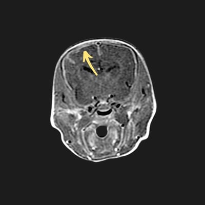
Post Contrast MRI of Brain:
Arrow points to white outline of brain indicating inflammation of the meninges

koda-Backteria-square-4897bb538ee179f777037bfad75bba42-6efkwnhg2sj0

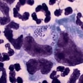
Spinal Fluid Analysis:
Increased number of inflammatory cells (longer purple areas) with bacteria present (small purple dots in center)

Medical Treatment

Bacterial meningitis is an uncommon, extremely serious, life-threatening condition; and Koda was severely affected.

She was hospitalized at SEVN and received IV antibiotic treatment and supportive care. At first, there was only minimal improvement, and we were quite worried whether this little girl would pull through or not. However, over the next few days, she made considerable improvement. Her white blood cell count began to decline, her mentation (mental activity) improved significantly, her nystagmus and strabismus improved, she began to eat and drink, and although wobbly, she began walking again.

She was transitioned to oral antibiotics and discharged into her owners’ care with the understanding that the full extent of her recovery could take time, and that some dogs would retain a degree of neurologic deficit.

After a long course of oral antibiotics at home, Koda continued to improve, and medications were discontinued. In July, Koda presented to SEVN for her last recheck and had a normal neurologic exam (save a subtle head tilt) and normal blood work. She was a happy, energetic, and otherwise healthy dog, and it was reported that she was doing very well at home. Koda made a dramatic recovery!

Takeaway

Meningitis is an inflammation of the membranes that cover and protect the brain and spinal cord. Inflammation in dogs is most commonly immune-mediated, but sometimes it can be infectious. Although not as common in dogs, infections may reach the central nervous system through the sinuses, inner ear, vertebrae, bloodstream, or even traumatic injuries.

The most important tests for diagnosing meningitis and suggesting an underlying cause are MRI and CSF analysis. This is important because the appropriate treatment and prognosis depend on the underlying cause. However, prognosis is generally guarded because relapses and death are common, even with appropriate treatment.

Veterinary neurologists have experience and expertise in neurological conditions, including uncommon conditions such as bacterial meningitis. Early, accurate, and aggressive treatment are critical to a positive outcome, and it doesn’t hurt to have a dedicated family, such as Koda’s! By coming to Southeast Veterinary Neurology and receiving a timely diagnosis and appropriate treatment, Koda’s family saved her life.

kodas-Graduation
Scroll To Top